An Everyday DNA blog article
Written by: Sarah Sharman, PhD, Science writer
Illustrated by: Cathleen Shaw
Parents in the United States in the 1940s and the 1950s lived in a state of fear for their children’s health because a highly infectious disease was preying on the young. The disease I am talking about is polio, which is caused by a family of viruses known as poliovirus. It infects a person’s spinal cord, causing symptoms like meningitis and paralysis. During the height of the U.S. polio epidemic of the 1940s and 1950s, polio was disabling more than 35,000 people each year.
Similar to what we’re seeing during the Covid-19 pandemic, scientists worked around the clock to develop a polio vaccine. At the time, viruses for research were grown in live animals or in animal products like chicken eggs. However, these methods could not quickly and affordably create enough virus to develop large quantities of polio vaccines.
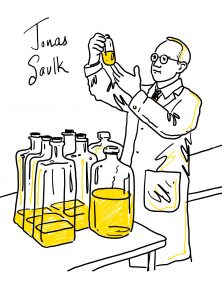
Enter a laboratory technique called animal cell culture. By growing billions of virus particles on monkey kidney cells, scientist Dr. Jonas Salk was able to create the first inactivated polio vaccine that became a major player in the efforts to eradicate poliovirus in the U.S. Let’s learn more about this valuable laboratory technique that has helped advance our knowledge of the inner workings of cells, develop life-saving vaccines and therapeutics, and bring us closer to understanding complex diseases like autoimmune diseases, cancer, and heart disease.
What is cell culture?
Cell culture is the process of removing cells from an animal or plant and growing them in a controlled laboratory setting where they can be used for experiments. For example, in the case of the polio vaccine, cells were removed from a monkey kidney, grown in Dr. Salk’s lab, and infected with poliovirus.
The origins of cell culture can be traced to the late 19th and early 20th centuries. In 1884, German zoologist Dr. Wilhelm Roulx demonstrated that living cells could be maintained outside of an organism by keeping embryonic cells from a chicken alive in saline for several days.
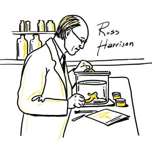
American biologist Dr. Ross Harrison is credited as being the “father” of animal cell culture as we know it today. In 1907, Harrison placed frog nerve cells in a droplet of lymph and observed the growth of nerve fibers for several weeks.
What followed was several decades of trial and error while the scientific community began to determine the best parameters and methods for successful cell culture. In order for cells to survive and multiply in cell culture, it is essential that the culture environment closely replicates the cells’ normal environment. While the lymph that Dr. Harrison used in his experiments provided the cells with enough nutrients to survive for several weeks, it was not a good long-term solution.
Thanks to the hard work of many scientists in the 1900s, scientists today grow their cells in a liquid called cell culture media. It contains nutrients to feed the cells and helps regulate pH and other factors to maintain a satisfactory environment for the cells. Different types of media are commercially available for many commonly used cell lines. Individual labs also make their own specific media for some primary cell lines.
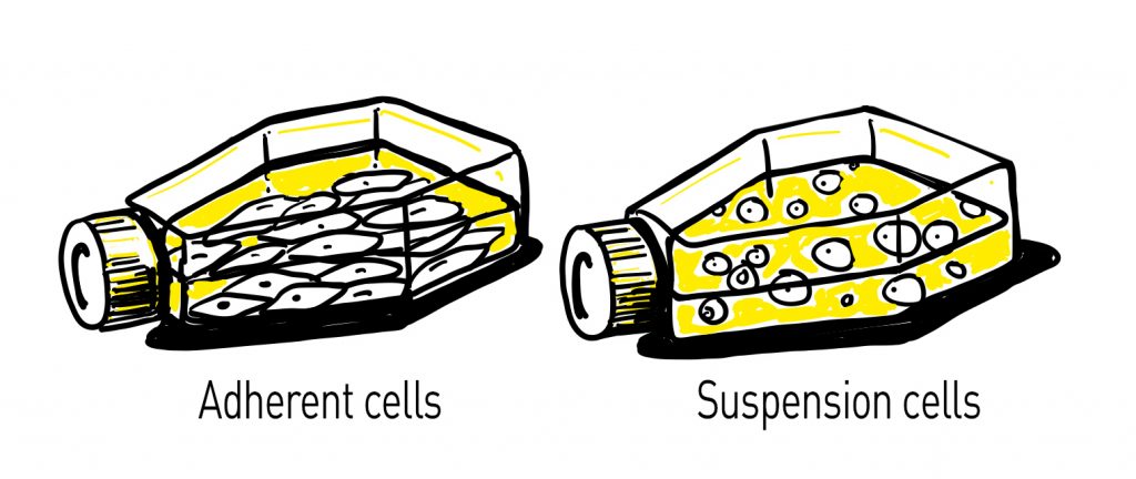
The cell culture media with the cells is usually contained in plastic dishes or flasks. Some cells called adherent cells stick to the bottom of the flask while others called suspension cells float freely in the media.
In addition to ample nutrients, cells also need to be maintained in a specific atmosphere. A piece of equipment called a cell culture incubator creates the perfect environment for cells. It resembles a refrigerator, but instead of keeping its contents cold, it maintains a warm temperature (around 37°C for mammalian cells) and appropriate humidity and (CO 2) levels for each cell type.
Speaking of cell types, you might be wondering what kind of cells scientists can grow in a lab. Both Dr. Roulx and Dr. Harrison were using what we call primary cell types in their early experiments. Primary cells are taken directly from an organism’s tissue without modifications. Examples of primary cells include epithelial cells, fibroblasts, endothelial cells, and muscle cells. Primary cells most closely resemble the normal state of a cell, however, they do have a limited life span.

As cell culture became more commonplace, scientists recognized that some cells called continuous cells, or immortalized cells, acquire the ability to reproduce indefinitely. Cells can become immortalized through random mutation (like transformed cancer cell lines) or deliberate modifications in a lab such as the expression of a cancer gene. Continuous cell lines have unlimited growth potential and grow quickly, however, they are not as biologically relevant as primary cells.
Within a cell culture suite in a research lab, you will also find a few more pieces of equipment that help cell-based experiments run smoothly and accurately. It is crucial to keep the cell culture working environment very clean to avoid contamination by other biological organisms, like bacteria and fungi. Most cell culture work is done within a cabinet-like piece of equipment called a cell culture hood. It features a downward flow of filtered air at its opening to reduce the likelihood of airborne contamination.
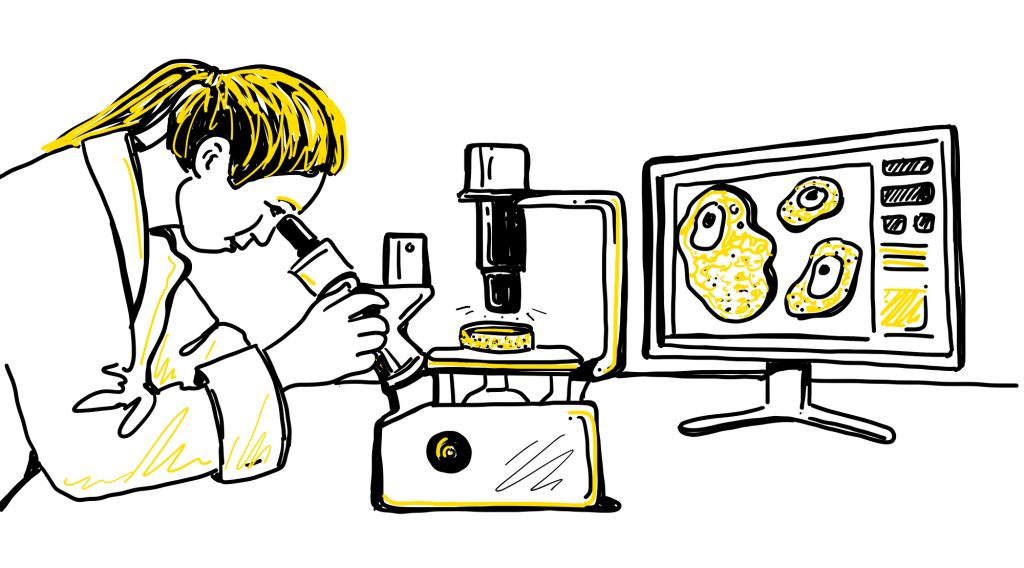
It is also common to keep microscopes within a cell culture suite. Microscopes help scientists visualize cells to examine cell growth and general cell health. Scientists often use dyes and stains to look at proteins and organelles within cells. Microscopes allow scientists to see the dyes within the cells.
What are the applications of cell culture?
Today, cell culture is a mainstay in research labs across the biological sciences and biotechnology industry. It has been one of the most important tools to allow scientists to learn the inner workings of cells. Many cell lines are used as model systems to study basic cell biology and biochemistry. They allow scientists to study the interactions between cells and disease-causing organisms like bacteria and viruses.
As we mentioned above with the polio vaccine, cell cultures can be used to produce large quantities of viruses for the production of vaccines. Vaccines for many harmful diseases like polio, rabies, chickenpox, measles, and hepatitis B are produced using cell culture. In addition to creating vaccines, cell culture can also be used to detect and isolate viruses and study the growth and development cycle of viruses.
Cell cultures are also used to produce commercially important genetically engineered proteins such as monoclonal antibodies, insulin, hormones, and more. They are frequently used in many steps of drug development from looking at the effects of the drug to drug screening to toxicity testing.
The basic difference between healthy cells and diseased cells can be studied using cell culture. Scientists can look at the differences between the cells to determine the cause of a disease or the mechanism of disease progression. They can also use cell culture to determine effective drugs for treating a disease.
At the HudsonAlpha Institute for Biotechnology, cell culture plays an important role in much of its human health research. A new cell culture suite was recently built to help support these growing research programs. The new suite includes much of the specialized equipment necessary for growing cells in culture, including cell culture hoods, incubators, centrifuges, and microscopes.
One research area that will rely heavily on the new space is the Memory and Mobility (M&M) Program led by Drs. Rick Myers and Nick Cochran. Their labs study several neurodegenerative diseases including Alzheimer’s, Huntington’s, Parkinson’s, ALS, and rare conditions such as frontotemporal dementia (FTD) and corticobasal syndrome (CBS).
The research teams use cell culture as a tool to study the genetic causes of neurodegenerative diseases, develop sophisticated blood-based biomarkers for early detection and disease monitoring, and discover new therapeutic approaches for these devastating diseases. The labs are also using a new technology called single-cell analysis to study neurodegenerative diseases on a cell by cell basis. Looking at individual cells provides the scientists with new, more precise information on the genetic differences between the brain of someone with a disease compared to someone without the disease.